

Organ and Body Scale WG
Development of HIFU simulator for non-invasive treatment with highintensity focused ultrasound
VCAD System Research Program, RIKEN
Kohei Okita
An ultrasonic imaging diagnostic system with which the inside of the body
can be investigated using inaudible ultrasound has been widely used in
clinical practice. There is a therapeutic method by which ultrasound stronger
than that used in this ultrasonic imaging diagnostic system is focused
on a target such as a tumor to necrotize tissues by heating, which is referred
to as HIFU (High Intensity Focused Ultrasound). The main characteristic
of HIFU is that treatment can be performed without incision, and the low
impact on the body is of great advantage. The treatment of uterine fibroids
and prostatic hyperplasia with therapeutic equipment using this HIFU has
already been approved in other countries, and clinical studies on the treatment
of liver tumors, etc., are being conduced and awaiting application for
approval, but some problems remain. In the treatment of a deep-seated liver
tumor, for example, the ultrasound transmitted from the HIFU system runs
from the skin through organs such as fat, muscles, bones and liver to a
focal point (right side of Figure 1). At this time, the ultrasound may
be absorbed so that it decays when it passes through various organs, the
ultrasound may be refracted so that it bends, and part of the ultrasound
may be reflected. Thus, in treating deep-seated tumors with HIFU, the energy
required to heat the target may be insufficient due to decay of the ultrasound,
the focal point may be blurred because of reflection or refraction of the
ultrasound, and the position of the focal point may deviate from the target.
In order to control the HIFU system so that the ultrasound focuses on the
target, it is therefore necessary to know how the ultrasound passes through
the body, and using the biological information obtained by CT or MRI, we
are trying to reproduce the behavior of ultrasound transmission in a biological
body by simulation.[1]
So far, the result of a simulation of HIFU treatment for a liver tumor
has been obtained as shown in Figure 1.[2] It is seen that the ultrasound
sent from the HIFU system is transmitted in a complex way as shown in Figure
2, and focuses short of the target. In such a case, the normal part is
heated instead of the site of tumor to be treated. In order to focus the
ultrasound onto the target by controlling the ultrasound delivered by the
system, a method called the time-reversal method[3] is therefore used.
The timereversal method is to emit ultrasound from a sound source placed
at the target point, to receive the ultrasound with the HIFU system, and
to send the received signals by reversing the time. The result of simulation
by controlling the HIFU system by this time-reversal method is shown in
Figure 3, showing that the ultrasound focuses appropriately on the target.
When ultrasound is transmitted in a complex way in the body as in this
case, it is expected that it will be possible to treat a deep-seated tumor
more precisely by controlling the HIFU system. Actually, since it is difficult
to place a sound source at the target depth in the body, it is possible
to perform highly precise HIFU treatment using the time-reversal method
by determining the controlling parameter of the HIFU system by simulation
in advance. Since a highly precise HIFU simulation is therefore required,
we will increase the precision of a HIFU simulator by verifying the simulation
result by comparison with experiment, and by introduction of a more advanced
physical model. For realization of a highly precise and non-invasive HIFU
treatment in the near future, we will contribute to the design and control
of a HIFU system, and clinical studies for approval and examination of
a preoperative treatment plan, etc., using the HIFU simulator.
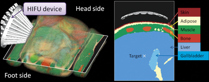
Figure 1 : HIFU simulation of liver tumor using a numerical human body model
The figure on the rights shows the distribution of tissues lying in the path of ultrasound
transmission.
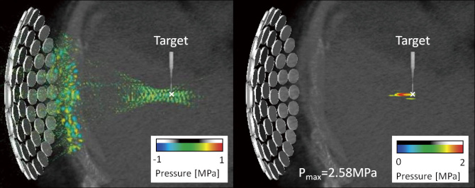
Figure 2 : Condition of ultrasound transmission (left) and the position of focal point (right)
when the HIFU system is not controlled
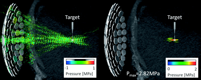
Figure 3 : Condition of ultrasound transmission (left) and the position of focal point (right)
when the HIFU system is controlled by the time-reversal method
References
1. Okita K., Ono K., Takagi S., Matsumoto Y., “Development of High Intensity Focused Ultrasound
Simulator for Large Scale Computing,” Int. J. Numer. Meth. Fluids, Vol. 65, pp. 43-66, 2011.
2. Okita K., Ono K., Takagi S., Matsumoto Y., “Numerical Simulation of the Tissue Ablation in High
Intensity Focused Ultrasound Therapy with Array Transducer,” Int. J. Numer. Meth. Fluids , Vol.
64, pp. 1395-1411, 2010.
3. Fink M., Montaldo G., Tanter M., “Time-reversal acoustics in biomedical engineering,” Annu.
Rev. Biomed. Eng ., Vol. 5, pp. 456-497, 2003.
BioSupercomputing Newsletter Vol.4
- SPECIAL INTERVIEW
- In order to change from observation-type medical practice focusing on experience to prediction-type medical practice to construct the base of theoretical medicine
Professor, Department of Internal Medicine (Cardiovascular Medicine), Director of the Metabolic Disease Research Center, Bio-Research Medical Center, Tokai University Graduate School of Medicine, and Director, Department of Metabolic System Medicine, Tokai University General Medical Laboratory Shinya Goto - It is expected that new possibilities in nutrition science and health control will be opened up
by simulation science
EXECUTIVE PROFESSIONAL Health infomatics DEPT., Ajinomoto Co., Inc. Toshihiko Ando
- Report on Research
- The functions of a multidrug discharging transporter were verified by coarse graining molecular simulation (Molecular Scale WG)
Graduate School of Science, Kyoto University Shoji Takada / Xin-Qiu Yao / Hiroo Kenzaki - Cell simulation considering time-space (Cell Scale WG)
Computational Science Research Program, RIKEN Yasuhiro Sunaga - Development of HIFU simulator for non-invasive treatment with high-intensity focused ultrasound (Organ and Body Scale WG)
VCAD System Research Program, RIKEN Kohei Okita - PLATO: Platform for a collaborative brain system modeling toward development of large scale mathematical model.(Brain and Neural WG)
①Computational Science Research Program, RIKEN
②Brain Science Institute, RIKEN
Keiichiro Inagaki①/ Takayuki Kannon②/ Nilton L. Kamiji②/ Koji Makimura②/ Shiro Usui ①②
- Report
- Report on the workshop in BMB2011 (Joint Meeting of the 33rd Congress of the Molecular Biology Society of Japan and the 83th Congress of the Japanese Biochemical Society)
- Winter School 2011 for the Integrated Simulation of Living Matter
Computational Science Research Program, RIKEN Yasuhiro Ishimine (Organ and Body Scale WG)
The Institute of Medical Science, The University of Tokyo Hidetoshi Urakubo (Brain and Neural WG)
Computational Science Research Program, RIKEN Yasuhiro Sunaga (Cell Scale WG)
Computational Science Research Program, RIKEN Gen Masumoto (High-Performance Computing Team)
Computational Science Research Program, RIKEN Keiji Misawa (Data Analysis Fusion WG)
Computational Science Research Program, RIKEN Hisayuki Miyashita (Molecular Scale WG) - Winter School 2011 for the Integrated Simulation of Living Matter