

Molecular Scale WG
The functions of a multidrug
discharging transporter were
verified by coarse graining
molecular simulation
Graduate School of Science, Kyoto University
Shoji Takada, Xin-Qiu Yao, and Hiroo Kenzaki
Multidrug resistance where most drugs become ineffective leads to serious
social problems in nosocomial infection and cancer chemotherapy, etc.
This multidrug resistance arises due to different mechanisms. In the case
of Pseudomonas aeruginosa which was a large problem in nosocomial
infection, the main cause of multidrug resistance was that expression
of a multidrug discharging transporter of RND type in Pseudomonas
aeruginosa increases and discharges antibiotics from the bacteria. The
multidrug discharging transporter of RND type is driven by transfer of
H+ (proton) using the difference in acidity (pH) inside and outside cells,
and discharges the drug by using this ability. The atomic structure of the
Escherichia coli-derived RND type multidrug discharging transporter
“AcrB” was elucidated by Mr. Satoshi Murakami (presently Professor of
Tokyo Institute of Technology), et al. using X-ray crystal structural analysis
in 2002 and 2006. In the structural analysis done in 2002, it was shown that
AcrB is a trimer of 3 similar molecules, having three-fold symmetry, while
in the structural analysis in 2006, it was found that each molecule has the
function of a membrane proton transporter and drug discharger, and that
the trimer of AcrB has an asymmetric structure. In the first molecule of this
asymmetric AcrB trimer structure, a route considered as an entrance for the
drug facing into the cell opens (incorporation type), in the second molecule,
the drug binds to the center (binding type), and in the third molecule, a
drug discharge port facing outward from the cell opens (discharging type).
Murakami et al. considered that the 3 molecules of the AcrB trimer discharge
drugs by mediating these 3 functional states in turn. Since it seems that the
whole structure rotates 120 degrees by changing the respective states of the
3 molecules step by step, this mechanism of drug discharge was named a
“functional rotation mechanism.” However, since the verification experiment
using this experimental system was difficult, it was impossible to verify this
hypothesis.
We have independently developed a technique for coarse graining
molecular simulation of biomolecules as part of the project “Research and
Development of Next-Generation Living Matter Integrated Simulation
Software” of the Ministry of Education, Culture, Sports, Science and
Technology. In this research, we conducted
a functional simulation attributable to
fluctuation of the multidrug discharge
transporter AcrB by applying this new
technique.
(1) Functional rotation of AcrB trimer and drug discharge: In the asymmetric AcrB trimer structure, when a proton binds to the drug-bound AcrB molecule (blue on the left side of Figure 1) from the extracellular space, the drug is discharged outward (center of Figure 1), and subsequently, the other 2 AcrB molecules also changed their state, and a functional rotation occurred (right side of Figure 1). By this, it was shown that functional rotation occurred according to proton binding and that drug discharge could occur.
(2) Resting state of AcrB trimer: It was found that, if a drug is removed from the asymmetric AcrB trimer structure, the structure having three-fold symmetry becomes stable. This structure is near the structure elucidated in 2002. That is, the structure obtained in the structural analysis in 2006 is a snapshot of the way the AcrB trimer discharges the drug, and the structure in 2002 is considered to correspond to the resting state.
Reference
Xin-Qiu Yao, Hiroo Kenzaki, Satoshi Murakami & Shoji Takada,
Drug export and allosteric coupling in a multidrug transpor ter revealed
by molecular simulations “Nature Communications. 1, 117 (8 pages) (2010)
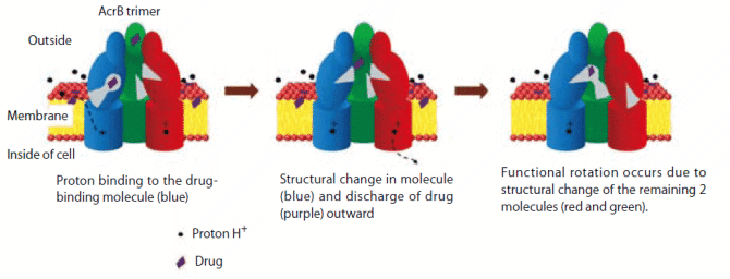
Figure 1 : Drug discharge and functional rotation of AcrB due to proton binding The AcrB trimer is of asymmetric structure. In the first molecule, the route considered as an entrance for the drug, which faces into the cell, opens (green in the left figure: incorporation type), in the second molecule, the drug binds to the center (blue in the left figure: binding type), and in the third molecule, the drug discharge port facing outward opens (red in the left figure: discharging type). When a proton binds to the second drug-binding molecule from the outside of a cell (arrow of dotted line in the left figure), the drug of this molecule is discharged to the outside of the cell (figure at the center), and the other 2 molecules change their states (right figure). |
BioSupercomputing Newsletter Vol.4
- SPECIAL INTERVIEW
- In order to change from observation-type medical practice focusing on experience to prediction-type medical practice to construct the base of theoretical medicine
Professor, Department of Internal Medicine (Cardiovascular Medicine), Director of the Metabolic Disease Research Center, Bio-Research Medical Center, Tokai University Graduate School of Medicine, and Director, Department of Metabolic System Medicine, Tokai University General Medical Laboratory Shinya Goto - It is expected that new possibilities in nutrition science and health control will be opened up
by simulation science
EXECUTIVE PROFESSIONAL Health infomatics DEPT., Ajinomoto Co., Inc. Toshihiko Ando
- Report on Research
- The functions of a multidrug discharging transporter were verified by coarse graining molecular simulation (Molecular Scale WG)
Graduate School of Science, Kyoto University Shoji Takada / Xin-Qiu Yao / Hiroo Kenzaki - Cell simulation considering time-space (Cell Scale WG)
Computational Science Research Program, RIKEN Yasuhiro Sunaga - Development of HIFU simulator for non-invasive treatment with high-intensity focused ultrasound (Organ and Body Scale WG)
VCAD System Research Program, RIKEN Kohei Okita - PLATO: Platform for a collaborative brain system modeling toward development of large scale mathematical model.(Brain and Neural WG)
①Computational Science Research Program, RIKEN
②Brain Science Institute, RIKEN
Keiichiro Inagaki①/ Takayuki Kannon②/ Nilton L. Kamiji②/ Koji Makimura②/ Shiro Usui ①②
- Report
- Report on the workshop in BMB2011 (Joint Meeting of the 33rd Congress of the Molecular Biology Society of Japan and the 83th Congress of the Japanese Biochemical Society)
- Winter School 2011 for the Integrated Simulation of Living Matter
Computational Science Research Program, RIKEN Yasuhiro Ishimine (Organ and Body Scale WG)
The Institute of Medical Science, The University of Tokyo Hidetoshi Urakubo (Brain and Neural WG)
Computational Science Research Program, RIKEN Yasuhiro Sunaga (Cell Scale WG)
Computational Science Research Program, RIKEN Gen Masumoto (High-Performance Computing Team)
Computational Science Research Program, RIKEN Keiji Misawa (Data Analysis Fusion WG)
Computational Science Research Program, RIKEN Hisayuki Miyashita (Molecular Scale WG) - Winter School 2011 for the Integrated Simulation of Living Matter